
Figure 7. Senescence of BMDCs in the postpartum uterus.
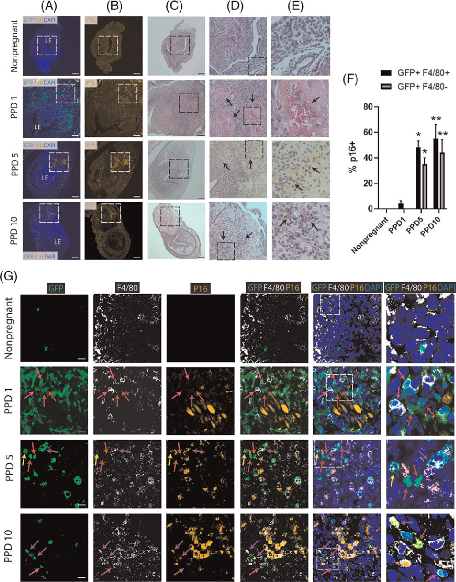
(A-B) Immunofluorescence of uterine tissue sections showing colocalization of GFP-positive BMDCs (green) and P16-positive senescent cells (orange) across postpartum time points and nonpregnant uterus; sections were counterstained with DAPI for nuclear staining (blue). ‘LE’ indicates the luminal epithelium. Note the white dashed squares on PPD1, PPD5 and PPD10 that are rich in p16-positive senescent areas within the implantation scar. The white dashed area in (B) is corresponding to the black dashed area in (C). (C-E) H&E staining of uterine tissue sections across postpartum time points and nonpregnant uterus. (C) 10x magnification showing black dashed square area which corresponds to the white dashed area in (A-B). (D-E) Column D are magnified (20x) H&E images of the black square in (C), and column E are magnified (40x) images of the black square area in (D). Black arrows on PPD1 in (D) are pointing to areas of fibrin deposition to the extracellular matrix and hemorrhage from blood vessel in (E). On PPD5 and PPD10, black arrows in (D) are pointing to areas rich in hemosiderin, a red blood cell product, indicating active areas of tissue organization and repair. Higher magnification images in (E) from PPD5 and PPD10 show black arrows pointing to hemosiderin-laden macrophages. Scale bar, 200 μm. (F) Quantitative summary of immunostaining from (G) showing percentage of GFP+/F4/80+ cells and GFP+/F4/80− cells that express p16 senescence marker across postpartum time points and nonpregnant state. n = 3 in all groups. * P < 0.05, and ** P < 0.01 vs nonpregnant and PPD1 timepoints.
(G) Immunofluorescence of uterine tissue sections from nonpregnant and across postpartum time points showing colocalization of GFP-positive BMDCs (green), F4/80 macrophage marker (white), and p16-positive senescent cells (orange). Sections were counterstained with DAPI for nuclear staining (blue). Red arrows point to nonhematopoietic GFP+ BMDCs that are negative for p16. Pink arrows point to GFP+/F4/80+ BM-derived macrophages that are p16-negative. Yellow arrows point to nonhematopoietic GFP+/P16+ senescent cells. Purple arrows point to GFP +/F4/80+/P16+ BM-derived senescent macrophages. Dashed white squares in merged images show tissue area that is magnified in the column to the right. Note the near absence of senescent (p16+) BMDCs on PPD1, and gradual appearance of p16+ BMDCs on PPD5 and PPD10. Scale bar, 20 μm