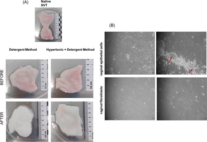
Figure 1.

(A) Gross appearance of the sheep vaginal tissue (SVT) before and after decellularization in two different solutions showing visible differences in appearance as the decellularized tissue is whiter in color and delicate upon handling with forceps compared to the native SVT. (B) Light microscopy images of primary sheep vaginal epithelial cells and fibroblasts isolated from sheep vagina under ×10 magnification. Epithelial cells showing cobblestone‐like appearance when cultured on irradiated mouse 3T3s (shown with red arrow heads surrounding the colonies). Vaginal fibroblasts showing flat, elongated morphology (scale bar = 100 μm)
