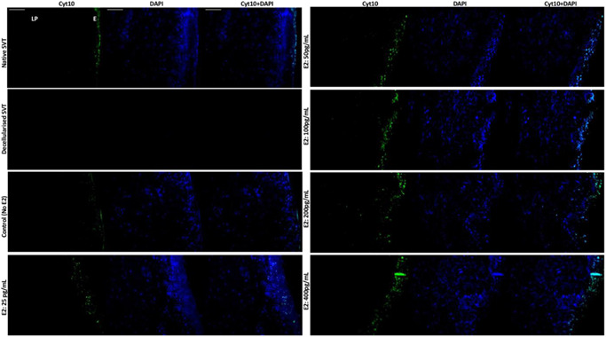
Figure 6.

Immunohistofluorescence detection of cytokeratin 10 (cyt10), a marker of stratification in the TE sheep vaginal models. The suprabasal cells and the superficial layers of the vaginal epithelium are stained intensely for cyt10 (green channel) in the native sheep vaginal tissue (top left), whereas no cellular expression for either cyt10 or DAPI could be seen in the decellularized sheep vaginal tissue (SVT) (negative control). In the reconstructed TE vaginal models, under estradiol‐17β [E2] induction, a dose‐dependent response of cells positive for cyt10 could be seen. Higher E2 concentration (from 50 to 400 pg/ml) showed an increase in the intensity and number of positive cells for the expression of cyt10 in the parabasal layers of the vaginal epithelium (E) while none of the vaginal fibroblasts in the lamina propria (LP) were positive for the cyt10 expression. All tissue sections were counterstained with DAPI (blue channel). Scale bar = 100 μm (applies to all). DAPI, 4′,6‐diamidino‐2‐phenylindole; TE, tissue engineered
