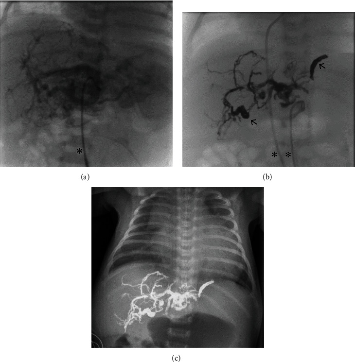
Figure 2.

Imaging of the hepatic AVM in patient 4. (a) Angiographical imaging after injection of Solutrast 300® into the feeding arteries via transarterial catheter (asterisk). (b) Angiographical imaging after injection of radiopaque Onyx® and Concerto Helix©-coils (arrows; two transvascular catheters are marked by asterisks). (c) Radiographic imaging (X-ray) of the thorax and upper abdomen after the last embolization, still existing extensive cardiomegaly.
