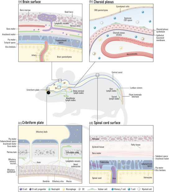
Fig. 1.

Overview of the brain barriers at select regions of the central nervous system (CNS). The mouse schematic displays the ventricular system (blue), comprised of two lateral ventricles (LV), the third ventricle (3V), the fourth ventricle (4V) and the central canal of the spine, and several known egress sites for cerebrospinal fluid (CSF) along the spinal and cranial nerves (yellow) to lymphatic vessels and draining lymph nodes (green). (a) Barriers at the dorsal surface of the brain cortex. The brain parenchyma is wrapped by the glia limitans, comprised of astrocyte end‐feet and an associated parenchymal basement membrane. Overlaying this is the pia mater meningeal layer, which along with the arachnoid mater encloses the subarachnoid space containing CSF and innate and adaptive immune cells performing CNS immune surveillance. The outermost layer of the meninges, the dura mater, contains venous sinuses draining the brain tissue, as well as its own blood vascular network (that does not form a blood–brain barrier) and dural lymphatic vessels. Peripheral immune cells thus have regular access to the dura mater. Channels surrounding blood vessels between the dura mater and bone marrow cavities may allow communication between these two compartments. (b) Blood–CSF barrier at the choroid plexus. The choroid plexus stroma is fed by a blood vessel network not endowed with barrier properties and harbors innate and adaptive immune cells. The barrier between this tissue and the CSF in the ventricles is formed by a layer of choroid plexus epithelial cells and an associated basement membrane. The CSF harbors low numbers of mainly adaptive immune cells performing CNS immune surveillance, as well as Kolmer (epiplexus) cells. The ventricle is lined by ependymal cells, which allows exchange of solutes between the CSF and the brain parenchyma. (c) CSF efflux routes at the cribriform plate. Olfactory nerve bundles extending from the olfactory bulbs project through foramina of the cribriform plate of the ethmoid bone to terminate at the epithelial layer of the nasal turbinates. The pia mater covering the olfactory bulbs blends into the perineurium surrounding the nerve bundles. At this region, the arachnoid layer is disrupted allowing passage of fluid, solutes (and possibly cells) out of the subarachnoid space. Lymphatic vessels, found either within the nasal mucosa or passing through the foramina along with the olfactory nerve bundles to the CNS side of the cribriform plate, are responsible for uptake and transport to the periphery. (d) Barriers at the surface of the dorsal spinal cord. The spinal cord parenchyma is wrapped by the glia limitans, comprised of astrocyte end‐feet and an associated parenchymal basement membrane. Veins exiting the spine are ensheathed by pia mater. The subarachnoid space lies between the pia mater and arachnoid mater and is traversed by trabeculae. It harbors innate and adaptive immune cells performing CNS immune surveillance. The spinal cord dura (unlike cranial dura) is associated with a subdural space and epidural tissue.
