Figure 2.

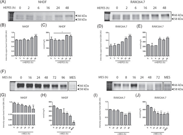
Induction and reversibility of GCase changes by HEPES‐containing medium. (A) Induction. Skin fibroblasts (NHDF) and RAW264.7 cells were exposed to either 50 mM HEPES or MES, and cellular GCase was monitored in time (0–48 h) by means of ABP labeling of enzyme in cell lysates and the measurement of enzymatic activity in lysates. (B) Quantified intensity of GCase glycan isoforms 62–66 in NHDF lysates depicted in (A), corrected for total GCase (total). (C) GCase activity of the same lysates of NHDFs was measured with 4‐MU‐β‐Glc substrate as described in Section 2. (D) Quantified intensity of GCase glycan isoforms 62‐66 in RAW264.7 lysates depicted in (A), corrected for total GCase (total). (E) GCase activity of the same lysates of RAW264.7 was measured with 4‐MU‐β‐Glc substrate as described in Section 2. (F) Reversibility. Skin fibroblasts (NHDF) and RAW264.7 cells were exposed for 3 days to 50 mM HEPES in the culture medium (pH 7.4). Following washing, cells were cultured in medium containing 50 mM MES (medium pH 7.0), and cellular GCase was monitored in time (0–96 h) in cell lysates by means of ABP labeling of enzyme molecules and measurement of GCase activity. (G) Quantified intensity of GCase glycan isoforms 62–66 in NHDF lysates depicted in (F), corrected for total GCase (total). (H) GCase activity of the same lysates of NHDFs was measured with 4‐MU‐β‐Glc substrate as described in Section 2. (I) Quantified intensity of GCase glycan isoforms 62–66 kDa in RAW264.7 lysates depicted in (F), corrected for total GCase (total). (J) GCase activity of the same lysates of RAW264.7 was measured with 4‐MU‐β‐Glc substrate as described in Section 2. The last lane to the right represents cells chronically cultured in in the presence of 50 mM MES. Overall significance of treatment effect (one‐way ANOVA, Tukey post hoc) is indicated by graph‐wide asterisks, individual asterisks on bars indicate significance compared to t = 0 (A) or HEPES (B). *p ≤ 0.05, **p ≤ 0.01, ***p ≤ 0.001. 4‐MU‐β‐Glc, 4‐methylumbelliferyl substrate beta‐d‐glucopyranoside; ABP, activity‐based probe; ANOVA, analysis of variance; HEPES, 4‐(2‐hydroxyethyl)‐1‐piperazineethanesulfonic acid; MES, 2‐(N‐morpholino)ethanesulfonic acid; NHDF, normal human dermal fibroblast
