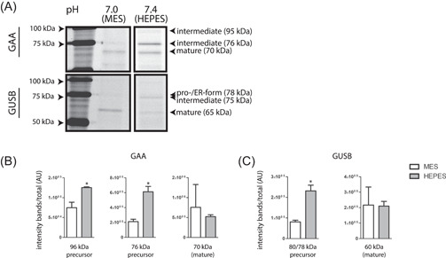
Figure 7.

Impact of medium pH on acid alpha‐glucosidase (GAA) and beta‐glucuronidase (GUSB) isoforms. (A) Fibroblasts were cultured in the presence of 50 mM buffer compound (MES or HEPES). Cells were harvested and GAA and GUSB in lysates was visualized by ABP labeling, SDS‐PAGE, and fluorescence scanning. (B) Quantified intensity of GAA isoforms depicted in (A), corrected for total GAA intensity. (C) Quantified intensity of GUSB isoforms depicted in (A), corrected for total GUSB intensity. ABP, activity‐based probes; HEPES, 4‐(2‐hydroxyethyl)‐1‐piperazineethanesulfonic acid; MES, 2‐(N‐morpholino)ethanesulfonic acid; SDS‐PAGE, sodium dodecyl sulfate‐polyacrylamide gel electrophoresis
