
FIGURE 1.
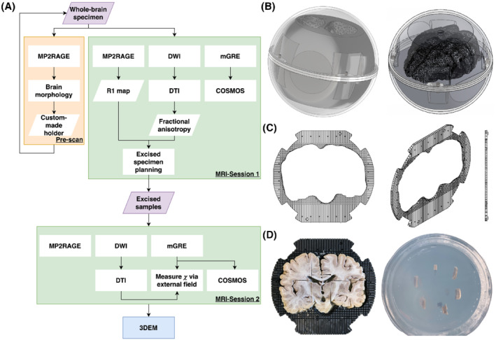
(A) Summary of this study consisting of a prescan, two MRI sessions, and a 3D electron microscopy (3DEM) session. (B) Schematic diagrams of the experimental setup used in the first MRI session. The setup is made of two parts: an outer transparent sphere that allows the specimen to rotate freely, and a tailor‐made inner holder to ensure the specimen was in a fixed position within the sphere. (C) Schematic diagrams of the plate that forms the inner holder. The center of the plate is a space with the shape of the specimen, surrounded by a grid structure providing a location reference in the MR image (the gaps were filled with water, whereas the plate material gave no detectable signal), and a guide during the sample excision for the second imaging session. (D) An illustration of how the samples were acquired with the aid of the plate (right) and were embedded in the agar inside the cylindrical container (left). Abbreviations: COSMOS, calculation of susceptibility through multiple orientation sampling; GRE, gradient echo