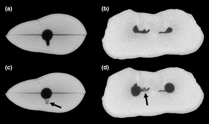
FIGURE 8.

Micro‐CT cross‐sections of extracted human teeth: a split and reassembled maxillary canine (a) before, and (c) after manual packing of dentine debris in an artificially‐created depression (arrow), and the mesial root of a mandibular molar (b) before, and (d) after preparation with rotary Ni‐Ti instruments. Note the dentine debris that accumulated in the uninstrumented fin of the molar during preparation (arrow)
