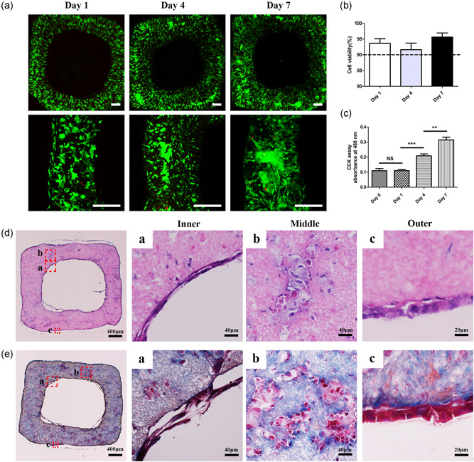
Figure 5.

Cell viability, proliferation, and histology of bionic vascular vessel. (a) Live/dead staining of bionic vascular vessel (Day 1, Day 4, and Day 7). Scale bar = 200 μm. (b) Cell viability of bionic vascular. (c) Cell proliferation by CCK‐8 assay (n = 4, **p < 0.01; ***p < 0.001). (d, e) The H&E staining and Masson staining. CCK‐8, Cell Counting Kit‐8; H&E, hematoxylin and eosin; NS, not significant
