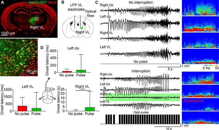
FIGURE 4.

Closed‐loop optogenetic inhibition of the ipsilateral VL does not change the onset latency in the contralateral VL or contralateral cortex. (A) ArchT‐GFP expressed in the right VL. (B) AAV9 CamKII‐ArchT‐GFP was injected in the right VL. (C) Seizures that were uninterrupted (no pulse) and interrupted (with opto pulse [black bars], green background in the right VL) in the same mouse with the corresponding power spectrums. (D) Seizure onset was delayed only in the right VL after pulse activation, whereas onset latency in the left cortex or left VL did not change. LFP = local field potential; VL = ventrolateral.
