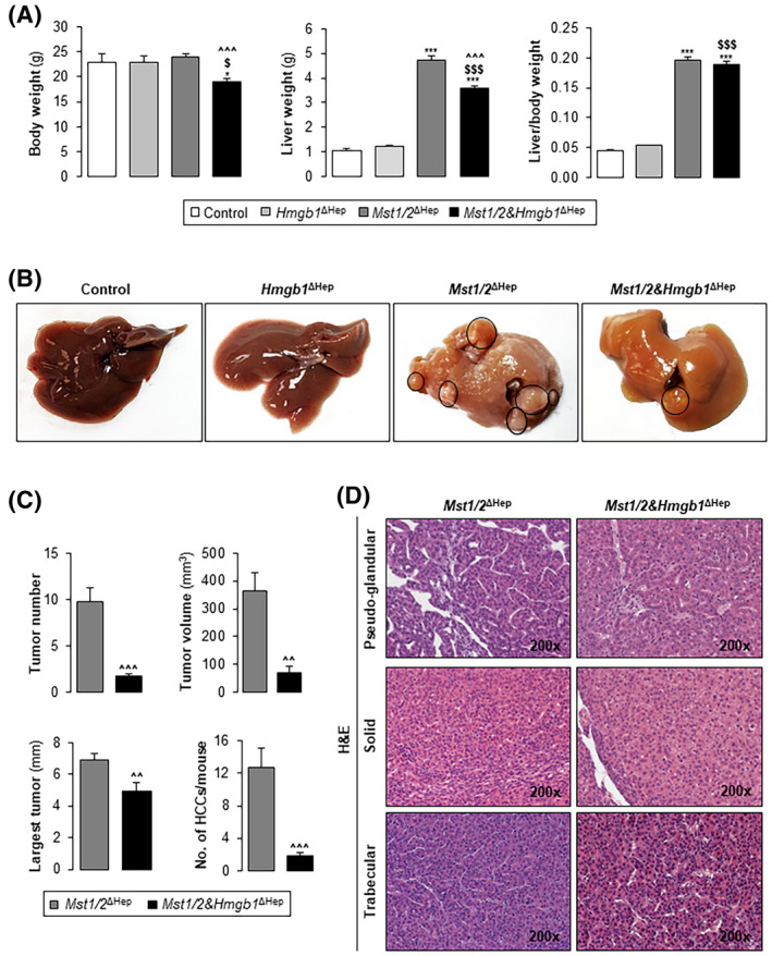
FIGURE 2.

Ablation of Hmgb1 in Mst1/2 ∆Hep mice reduces tumor burden. Control, Hmgb1 ∆Hep, Mst1/2 ∆Hep, and Mst1/2&Hmgb1 ∆Hep mice were generated and followed for the development of tumors for 3.5 months. (A) Body weight, liver weight, and liver‐to‐body weight ratio. (B) Macroscopic appearance of livers with tumors circled in black. (C) Tumor number, largest tumor diameter, total tumor volume, and number of HCCs/mouse. (D) H&E staining showing HCCs with pseudoglandular, solid, and trabecular growth patterns (control n = 6; Hmgb1 ∆Hep n = 5; Mst1/2 ∆Hep n = 12; Mst1/2&Hmgb1 ∆Hep n = 9). Values are given as mean ± SEM. *p < 0.05, ***p < 0.001 for Mst1/2&Hmgb1 ∆Hep or Mst1/2 ∆Hep versus control; $ p < 0.05 and $$$ p < 0.001 for Mst1/2&Hmgb1 ∆Hep versus Hmgb1 ∆Hep; ^^p < 0.01 and ^^^p < 0.001 for Mst1/2&Hmgb1 ∆Hep versus Mst1/2 ∆Hep. Abbreviations: HCC, hepatocellular carcinoma; H&E, hematoxylin and eosin; HMGB1, high‐mobility group box‐1; MST1/2, mammalian sterile 20‐like 1 and 2
