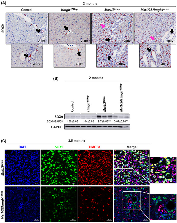
FIGURE 6.

Ablation of Hmgb1 in Mst1/2 ∆Hep mice reduces SOX9+ ADCs. Control, Hmgb1 ∆Hep, Mst1/2 ∆Hep, and Mst1/2&Hmgb1 ∆Hep mice were generated and followed for the development of tumors for 2 or 3.5 months. (A) SOX9 IHC at 2 months (black arrows show staining in anatomical BDs; pink arrows show staining in ADCs). (B) Western blot analysis of liver SOX9 at 2 months. (C) Immunofluorescence showing colocalization of DAPI (blue), SOX9 (green), and HMGB1 (red) (magnification ×630). Scale bar, 20 µm. Values are given as mean ± SEM. ***p < 0.001 for Mst1/2 ∆Hep versus control; ^^p < 0.01 for Mst1/2&Hmgb1 ∆Hep versus Mst1/2 ∆Hep. Abbreviations: ADC, atypical ductal cell; BD, bile duct; DAPI, 4′,6‐diamidino‐2‐phenylindole; GAPDH, glyceraldehyde 3‐phosphate dehydrogenase; HMGB1, high‐mobility group box‐1; IHC, immunohistochemistry; MST1/2, mammalian sterile 20‐like 1 and 2; Sox9, sex‐determining region Y‐box 9
