FIGURE 2.

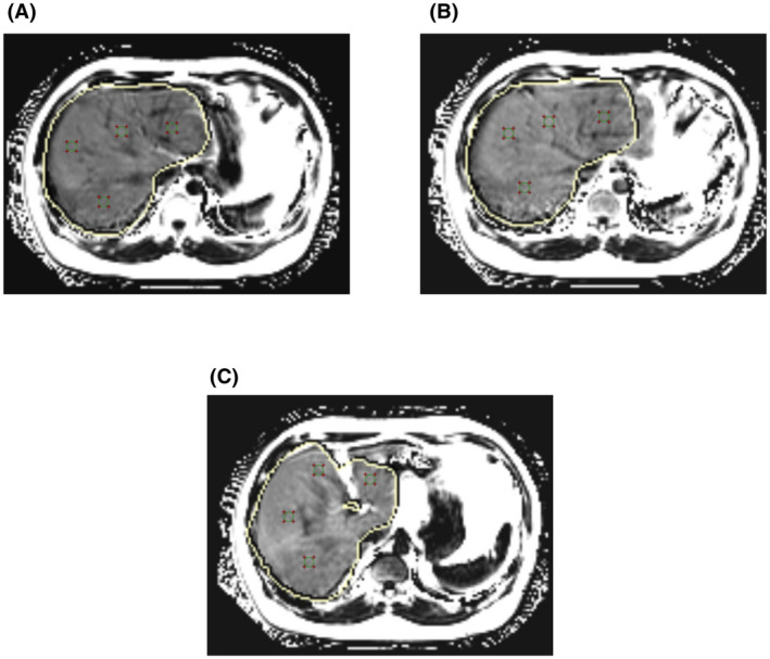
Representative MRI‐PDFF images of the liver of a 32‐year‐old man, with four regions of interest per slice. (A) First and (B) second hilar and (C) gallbladder fossa levels. All region of interest areas are 100 mm2

Representative MRI‐PDFF images of the liver of a 32‐year‐old man, with four regions of interest per slice. (A) First and (B) second hilar and (C) gallbladder fossa levels. All region of interest areas are 100 mm2