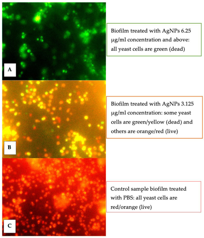
Figure 3.
Fluorescence microscope images of the C. auris viability assay using the Live/Dead yeast kit (Thermo Fisher Scientific, Waltham, MA, USA); red/orange indicates live cells; green/yellow indicates dead cells. The figure shows a noticeable variation in the cell viability of silver nanoparticle-treated biofilm. (A) Biofilm treated with 6.25 μg/mL showed green/yellow yeast cells (B) Biofilm treated with 3.125 μg/mL showed cells in green/yellow and orange/red (C) Untreated biofilm samples showed orange/red cells.

