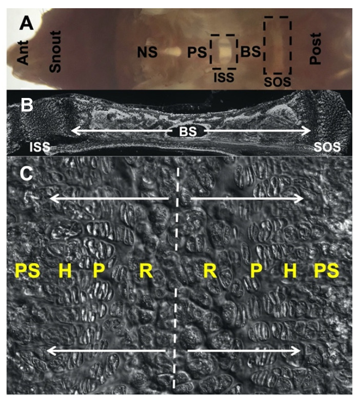
Figure 1.
Morphology of cranial base and spheno-occipital synchondrosis (SOS), C57BL/6 mouse at postnatal day 28. (A) Gross morphology of dissected cranial base positioned in an anteroposterior manner and taken from the dorsal view following the removal of the brain and cranial vault. (B) Sagittal section of inter-sphenoid synchondrosis (ISS), basisphenoid bone and SOS. (C) Magnified image highlighting bidirectional arrangement of chondrocyte layers in SOS. Ant: anterior, NS: nasal septum, PS: pre-sphenoid, BS: basisphenoid, Post: posterior, ISS: inter-sphenoid synchondrosis, SOS: spheno-occipital synchondrosis, R: resting zone, P: proliferating zone, H: hypertrophic zone, PS: primary spongiosa.

