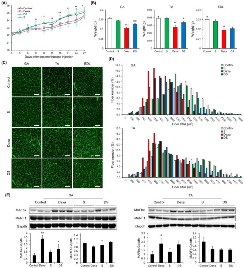
Figure 1.
β-sitosterol prevents Dexa-induced muscle atrophy in mice. (A) Body weight. * p < 0.05 and ** p < 0.01 control vs. Dexa. (B) Muscle weight of GA, TA, and EDL. ** p < 0.01 and *** p < 0.001 vs. control. # p < 0.05 and ### p < 0.001 vs. Dexa. (C) Representative immunofluorescent staining of myofiber cross section of GA, TA, and EDL. A microscope with a 10× objective was used to capture the images. The scale bar represents 100 μm. (D) Quantification of myofiber size by cross-sectional area (CSA) measurements for GA and TA muscle. Data are shown as mean ± S.E. (n = 9 per group). (E) Representative images of the western blot analyses for MAFbx and MuRF1 in GA and TA. # p < 0.05, ## p < 0.01 vs. control; * p < 0.05 vs. Dexa. GA—gastrocnemius muscles; TA—tibialis anterior; EDL—extensor digitorum longus; Dexa—Dexamethasone; S—β-sitosterol; DS—dexa + β-sitosterol.

