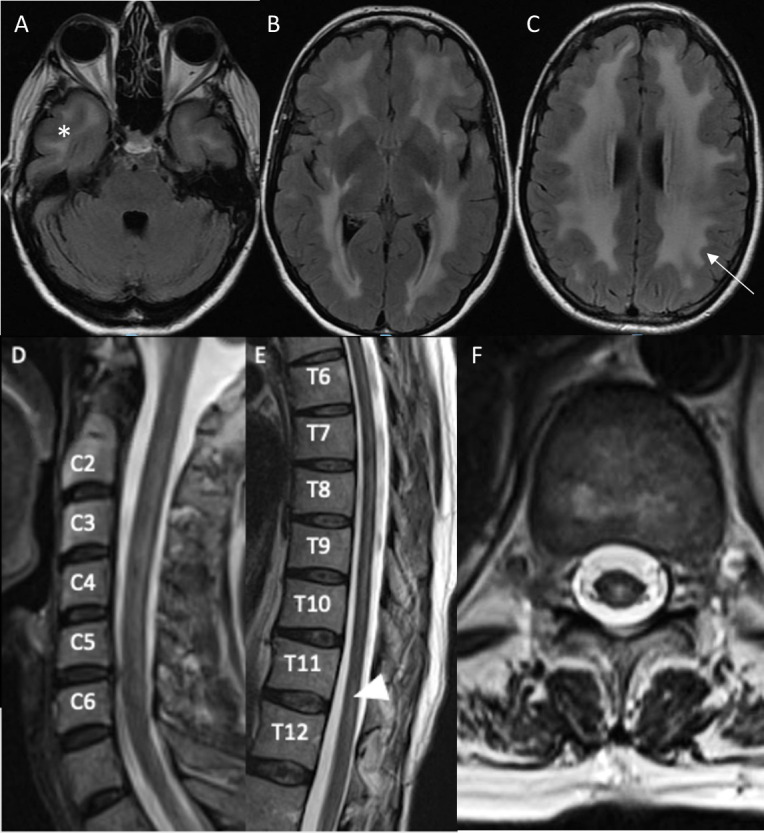
Figure 1.

MRI of the brain (panels A–C) 1 year prior to presentation and MRI of the cervical (panel D) and thoracic (panel E–F) spine at time of presentation. Post-gadolinium images are not shown as they were unremarkable. T2-weighted axial fluid-attenuated inversion recovery (FLAIR) images of the brain reveal symmetric confluent white matter hyperintensities in the bilateral anterior temporal lobes (A, asterisk), deep subcortical (B) periventricular and juxtacortical (C) regions of the brain with sparing of most of the occipital lobes and posterior fossa. The U fibres are mostly involved (C, arrow). The sagittal T2-weighted image of the cervical spine reveals a patchy extensive lesion most confluent from C3-5 (D). The sagittal thoracic spinal cord T2-weighted image reveals a longitudinally extensive hyperintense lesion most prominent from T4-T9 (partially seen in F) and a hyperintense lesion at T11-12 (E, F, arrow head) that are central dorsal in location, as corroborated on T2-weighted axial (F) image.
