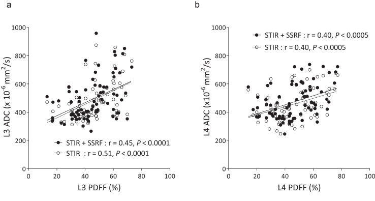
Fig. 4.
Correlation between lumbar PDFF and ADC in L3 (a) and L4 (b). Regression lines are separately shown with Spearman’s correlation coefficients r and associated P-values. ADC, apparent diffusion coefficient; DWI, diffusion-weighted imaging; SSRF, spectral-spatial RF; STIR, short inversion time inversion recovery.

