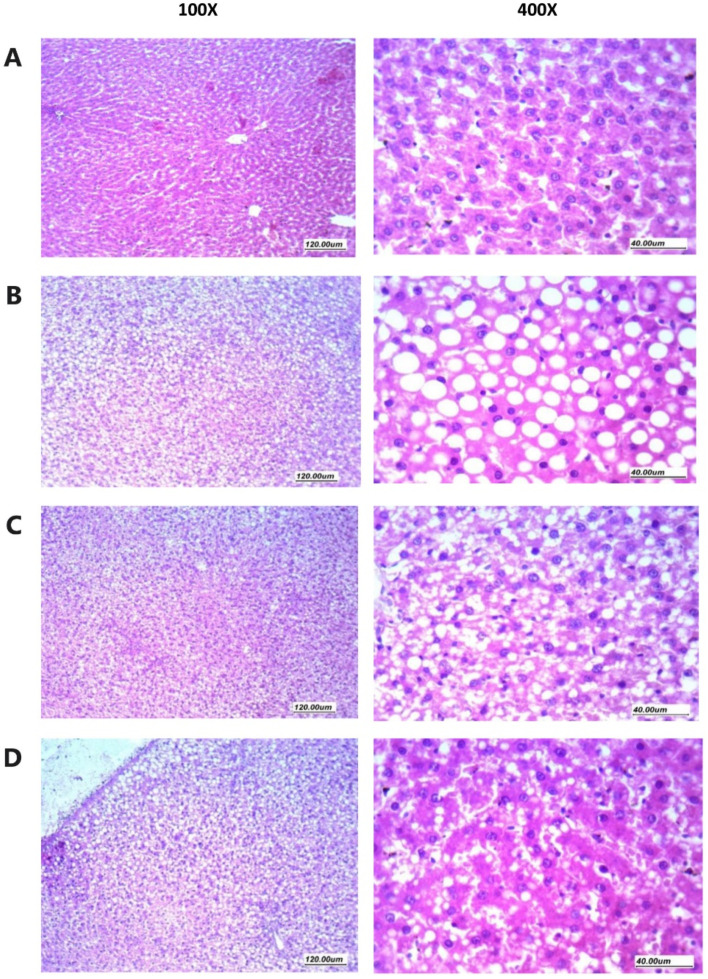
Figure 5.
Histopathology of rat liver specimens stained with hematoxylin and eosin. (A) Sections from normal liver tissues showing preserved architecture, hepatocytes arranged in thin plates separated by patent sinusoids. Hepatocytes have having homogenous eosinophilic cytoplasm, with regular nuclei. (B) Sections from liver tissues in NASH control group showing disturbed architecture, hepatocytes arranged in thick plates with marked steatosis (macro vesicular) presented as large cytoplasmic clear regular vacuole, with obscured sinusoids in-between. Nucleus is pushed to one side of the cells with regular or compressed contours. (C) Sections from liver tissues from NASH + hemp seed oil group showing moderately obscured sinusoids and predominantly preserved architecture, hepatocytes showing moderate steatosis (micro vesicular) with small to moderate-sized cytoplasmic clear vacuoles and regular vesicular nuclei. (D) Sections from liver tissues from the NASH + NEF#4 of hemp seed oil group showing predominantly preserved architecture, and patent sinusoids, hepatocytes arranged in thin plates, hepatocytes showing mild focal steatosis (micro vesicular) with a small number of scattered small-sized cytoplasmic clear vacuoles and regular vesicular nuclei.

