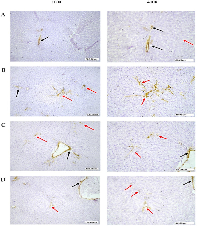
Figure 7.
Immunohistochemistry for alpha-smooth muscle actin in rat livers. (A) Normal liver tissue showing SMA staining limited to vascular walls (black arrow). The surrounding liver parenchyma shows no SMA staining (red arrow). (B) Liver tissues from NASH control group showing moderate SMA staining of stellate myofibroblast cells (red arrow) extending between liver cells within liver parenchyma with arborizing branching pattern between hepatocytes along sinusoidal walls. There are scattered vessels showing staining at periphery (black arrow). (C) Liver tissues from the NASH + hemp seed oil group showing focal moderate SMA staining of stellate myofibroblast cells (red arrow) extending early between liver cells within liver parenchyma. There are scattered vessels showing staining at the periphery (black arrow). (D) Liver tissues from the NASH + NEF#4 hemp seed oil group showing minimal very focal weak SMA staining of a very small number of myofibroblast cells (red arrow), minimally extending between liver cells within the liver parenchyma. There are scattered vessels showing staining at the periphery (black arrow).

