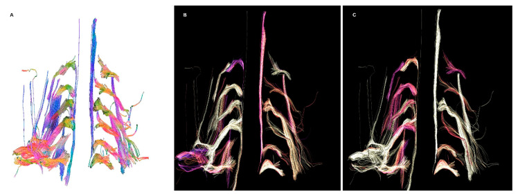
Figure 2.
(A) Fiber tracking showing an enlargement of the right brachial plexus, more marked in C5. (B) No significative differences in fractional anisotropy were noted, besides the cords of the right brachial plexus. (C) Decreased normalized quantitative anisotropy of the root and trunk of C5, and less evident of C6.

