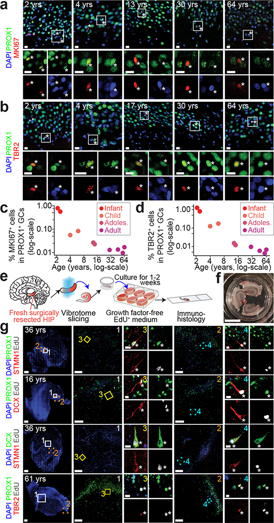
Fig. 5 |. Capacity for neurogenesis in the postnatal human hippocampus across ages.
a-d, Sample confocal immunostaining images (a, b) and quantifications (c, d) of PROX1+ neuronal progenitors in the human dentate gyrus across ages. Scale bars, 10 μm. Asterisks indicate MKI67+ or TBR2+ among PROX1+ GCs (a, b). Each dot represents the sum value of quantification of multiple sections from one specimen (n = 10 specimens) (c, d). e-g, A slice culture system demonstrating capacity for neurogenesis in the adult human dentate gyrus. Shown are a schematic illustration of the experimental procedure (e), a sample image of a well containing three slices (f; scale bar, 1 cm), and sample confocal staining images (g) of EdU-incorporated newborn imGCs expressing different markers in the postnatal human dentate gyrus. Scale bars, 100 μm (low-magnification images) and 10 μm (insets 3, 4) (g).

