
Fig. 4.
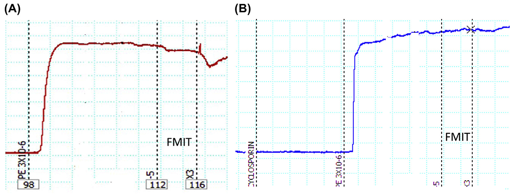
Representative tracing of formyl peptide mitochondrial (FMIT)-induced relaxation in resistance arteries (third order mesenteric arteries) from Wistar rats. The segments were initially contracted with phenylephrine (PE, 3 μM). Subsequently, FMIT (formyl–methionine–methionine–tyrosine–alanine–leucine–phenylalanine, 10 or 30 μM) was added in the absence or presence of cyclosporin H (FRP antagonist: CsH, 1 μM). As shown in (A), both concentrations of FMIT (10 or 30 μM) induced relaxation and the pre-incubation with CsH inhibited this response (B). Wire myograph was used to analyze vascular function.