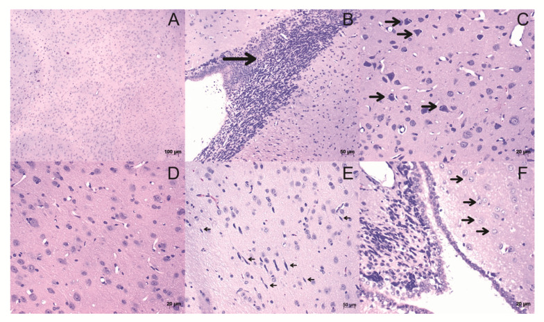
Figure 3.
Histological sections of brain stained with H&E. (A,D) Tissue from control animals, with a homogeneous distribution of brain cell types. (B) Leukocyte infiltrations (arrow). (C) Neurons on cell death: Observing neurons accompanied by leukocytes (arrows). (E) Activation of microglia (arrows). (F) Astrocyte activation (arrows). The images were obtained from optical microscopy (Carl Zeiss Microscopy, Zeiss, Jena, Germany). Scale bar, 50 µm.

