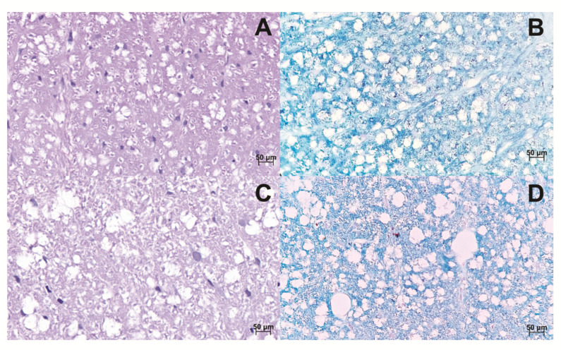
Figure 6.
Histological sections of the Spinal cord. The spinal cord of control animals was stained in HE and Luxol fast blue, respectively (A,B). The spinal cord of animals induced to the EAE model, stained in HE and Luxol fast blue, respectively. It is possible to observe the loosening of the myelinated fibers by increasing the axonal space (C,D). The images were obtained from optical microscopy (Carl Zeiss Microscopy, Zeiss, Jena, Germany). Scale bar, 50 µm.

