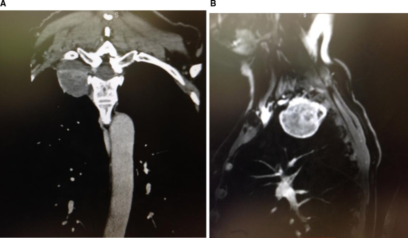
Figure 6.
Pre-operative coronal and sagittal thoracic MRI images showing high apical chest tumor. Thin slice CT images can be very helpful in identifying the neural foramen of tumor origin. This is critical in safely detaching the tumor from the spinal canal before final removal through the chest cavity.

