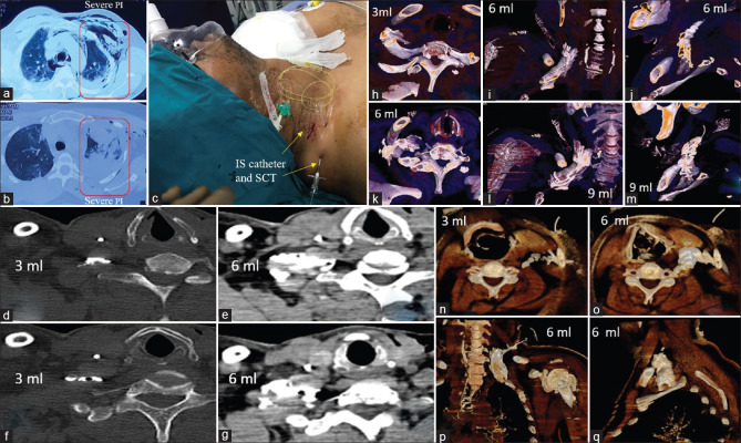
Figure 1.
(a and b) Right multiple rib fractures, subcutaneous emphysema, haemothorax (c) Right continuous interscalene block with subcutaneous tunnelling (d) Computed tomography axial 3 ml contrast in interscalene groove (e) Computed tomography axial 6ml contrast spilling over the scalene muscles (f and g) Computed tomography axial 3 ml sandwiched in interscalene groove (h) Axial volume rendering technique [inferior view] depicts linear contrast delineating the interscalene groove (hollow area).3 ml injection highlighting the middle scalene muscle and overlying the first rib (i) Coronal volume rendering technique image depicting the flow of 6 ml from medial to lateral and below the clavicle (j) Sagittal volume rendering technique image depicting the flow of 6 ml from medial to lateral and below the clavicle and in approximation to first rib (k) With 6ml injection, the inferior view portrays the three roots which appear distinctively, as the contrast spreads more laterally and posteriorly over the middle and posterior scalene muscles (l) Sagittal volume rendering technique image depicting the flow of 6ml from cephalad to caudal and between the clavicle and the first rib (m) Sagittal volume rendering technique image depicting the expansion as 9ml occupies the brachial plexus sheath (n) Axial volume rendering technique image; 3ml contrast is restricted to the interscalene groove (o) Axial volume rendering technique image illustrates spread over the anterior scalene muscle and beneath the sternocleidomastoid (p) Coronal volume rendering technique depicts the contrast spread in the interscalene groove and spilling over the scalene muscles (q) Sagittal volume rendering technique portrays the contrast as a thick band beneath the clavicle and close to the first rib. (IS – interscalene; SCT – subcutaneous tunnelling; 3 ml, 6 ml, 9 ml – volume of contrast injected; PI – pulmonary injury)

