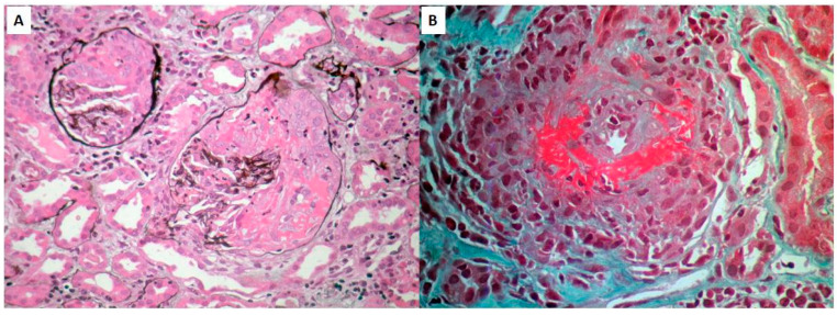
Figure 1.
PAS stain of renal biopsy showing two glomeruli with cellular crescents, covering >50% of glomerular area, complicated by necrosis within the crescents (A) and GT stain of an interlobular artery illustrating necrotizing vasculitis with lymphocytic infiltration and intimal proliferation, leading to marked elimination of the arterial lumen. Architectural changes and focal necrosis on the vessel wall are also prominent (B).

