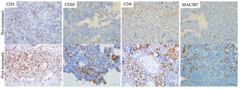
Figure 5.
Tumor Infiltrated immune cells. (brown) on formalin-fixed paraffin-embedded (FFPE) pre-(top) and post-treatment (bottom) biopsies including hematoxylin counterstaining (blue). T lymphocytes (CD3) of PSit02 at d0 and d332, T helper lymphocytes (CD4) of PSit06 at d0 and d126, B lymphocytes (CD20) of PSit02 at d0 and d332, and monocytes/macrophages (MAC387) in PSit06 at d0 and d28 are shown. Scale bar: 50 µm.

