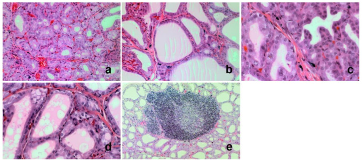
Figure 1.
Histopathology of selected rat thyroid glands: (a) Cuboidal follicular epithelial cells lining small follicles. Haematoxylin and Eosin (H + E) original magnification 200×. (b) Flattened follicular epithelial cells lining larger follicles. H + E original magnification 200×. (c) Few small papillary projections of follicular epithelial cells protruding into the lumina of the follicles. H + E original magnification 400×. (d) Vacuolation of the colloid. H + E original magnification 400×. (e) Lymphoid follicles. H + E original magnification 400×.

