Fig. 2.
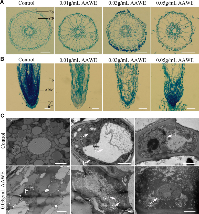
AAWE-treated rice plants have defects in the growth and development of root tip cells. A Microstructure of root tip cells by observing transection. Ep, epidermal layer; CP, cortical parenchyma; En, endothecium; P, pericycle. Scale bars = 100 μm. B Microstructure of root tip cells by observing longitudinal sections. Ep, epidermal layer; AM, apical meristem; QC, quiescent centre; RC, root cap. Scale bars = 200 μm. C Transmission electron microscopy of control and AAWE-treated root tip cells in rice. Two left panels, middle panels, and right panels indicate apical sections, single cells, and nuclei, respectively. 1, endoplasmic reticulum; 2, Golgi body; 3, vacuole; 4, nucleus; 5, fungi. Scale bars: 10 μm, 10 μm, and 1 μm (the first row runs from left to right); 10 μm, 1 μm, and 1 μm (the second row runs from left to right)

