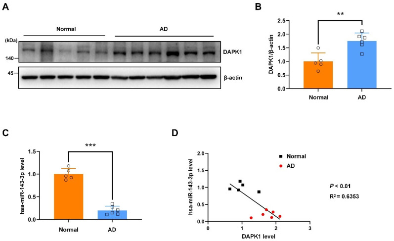
Figure 9.
The hsa-miR-143-3p levels are decreased and inversely correlated with DAPK1 expression in the hippocampus of AD patients. (A,B) Hippocampal tissues of AD patients and age-matched healthy controls were harvested, and the levels of DAPK1 were analyzed by immunoblotting. (C) The hsa-miR-143-3p levels in AD and normal control brain tissue samples were analyzed by qRT-PCR using U6 small nuclear RNA as an endogenous control. (D) Linear regression analysis was performed to evaluate the correlation between the DAPK1 and hsa-miR-143-3p levels (R2 = 0.6353; Pearson’s correlation coefficient). Relative quantification was performed using ImageJ software, and the results are presented as a histogram. The data (circles and squares) are presented as the mean ± SD of three independent experiments (** p < 0.01, *** p < 0.001).

