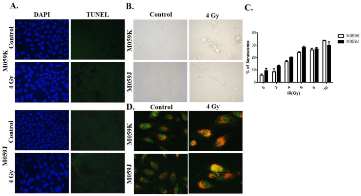
Figure 2.
Promotion of autophagy and absence of senescence and apoptosis upon exposure to radiation in M059K and M059J cells. (A). Cells were treated with a dose of 4 Gy and stained with DAPI (left) and TUNEL (right) to determine the portion of cells that undergo apoptosis upon radiation. Irradiated cells (bottom) images show no TUNEL-positive cells in both cell lines suggestive of minimal apoptosis induction. (B). SA-β-galactosidase upregulation was monitored in M059K and M059J cells to evaluate senescence markers post-radiation. Images show minimal expression of SA-β-galactosidase in both cell lines. (C). Quantification of senescence in M059K and M059J cells by flow cytometry-based measurement of the SA-β-galactosidase fluorogenic surrogate C12FDG. (D). Both cell lines were exposed to 4 Gy and stained with acridine orange 72 h post-treatment. Treated cells showed increased accumulation of acidic vacuoles and enlargement of cell morphology. All images were captured under 200x magnification.

