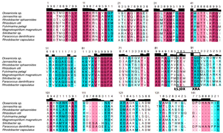
Figure 2.
Alignment of the ε subunits of the α-protebacterial ATP synthase. The sequences shown were aligned with Clustal Omega, and the alignment was visualized in UCSF Chimera. Color codes: cherry red: strongly conserved residues; pink, conserved residues; light pink, less conserved residues;  , slightly variable residues; light blue, variable residues; pale blue, average variable residues; strong blue, strongly variable residues. The most conserved region of the Pd-ε subunit is in the N-terminal side, the function of which is structurally connecting the γ subunit of F1 with the c12 ring of FO; see the case of the P. denitrificans F-ATP synthase in Figure 1C. The Pd-ε subunit is the largest one of this alignment, even larger than that of Oceanicola sp. with additional 9 a.a.s at the C-terminus. The C-terminus of α-proteobacterial ε subunit is highly variable, and this divergence leads to the loss of the ATP-binding motif of other bacterial inhibitory ε subunits (I(L)DXXRA) (see black box). The divergence of the C-termini of the α-proteobacterial ε subunits very likely produces the loss of both the inhibitory function of the two C-terminal α-helical hairpin domains and the loss of the regulatory ATP-sensor-binding motif. See text for further details.

