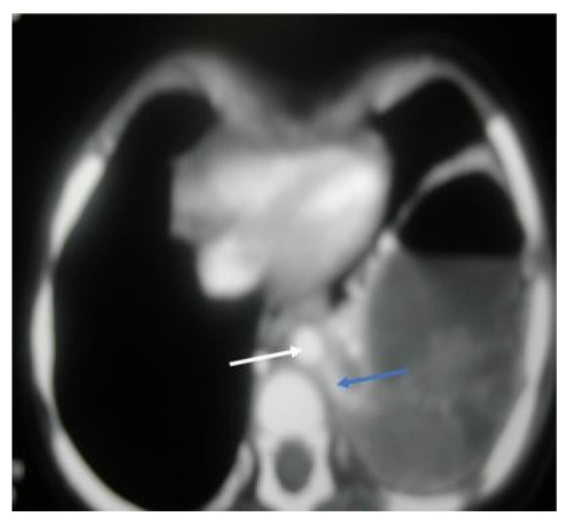
Figure 4.
CT chest, axial window, mediastinal view: large, thick wall cavity filled with air fluid with heterogenous opacity. The white arrow shows a descending aorta, while the blue arrow confirms a large feeding vessel to the left lower lobe, a rising and descending aorta, mild compression of left upper lobe, and a mild pectus excavatum.

