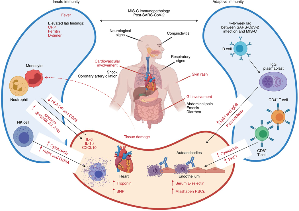
Fig. 2 |. Immunopathology associated with MIS-C post-SARS-CoV-2.
MIS-C is characterized by fever and cardiovascular, GI tract, neurological, respiratory and mucocutaneous inflammation. Immunological features include activation of both innate and adaptive responses, including elevated expression of genes encoding S100 alarmins and increased concentrations of acute phase proteins (CRP, ferritin and D-dimers) as well as IL-6, IL-1β and CXCL10, indicative of systemic inflammation. Activation of NK and CD8+ T cell cytotoxicity probably contributes to inflammation, as well as increased numbers of proliferating plasmablasts, which may produce autoreactive IgGs. Endothelial damage is evidenced by elevated concentrations of soluble E-selectin in the serum and cardiac damage by increased amounts of troponin and N-terminal pro-B-type natriuretic peptide (BNP). Figure adapted with permission from ref. 18, Cold Spring Harbor Laboratory; made in ©BioRender—biorender.com.

