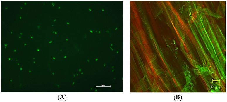
Figure 2.
(A) A bacterial suspension of GFP-tagged Pseuodomonas migulae 8R6 cells. Epifluorescence microscope, 100X objective lens. Photo courtesy of Patrizia Cesaro. (B) Bacterial cells of P. fluorescens 92GFPrk along the primary root of a 7-day-old tomato plantlet. Confocal Laser Scanning microscope, 100X objective lens.

