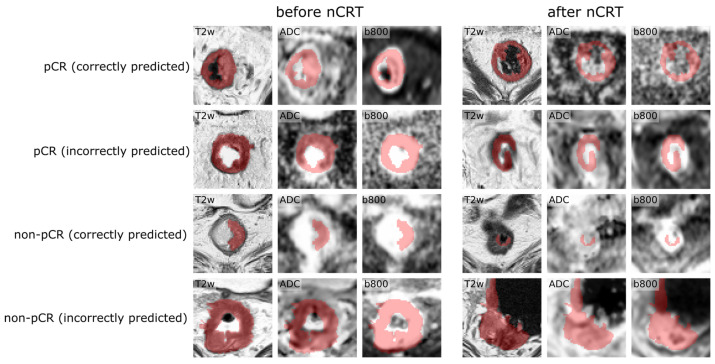
Figure 5.
Examples of correctly and incorrectly classified patients. Each row shows input images of one patient before and after nCRT overlayed with the segmentation mask. Note the slight misalignment of the segmentation mask with the tumor region on the DW images, which might contribute to misclassification.

