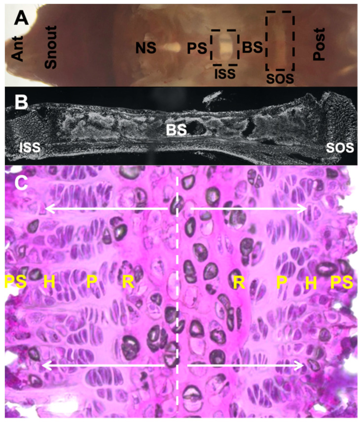
Figure 1.
Morphology of cranial base and SOS harvested at postnatal day 28 from C57BL/6 mouse. (A). Gross morphology of dissected cranial base positioned in an anteroposterior manner. (B). Sagittal section of ISS, basi-sphenoid bone and SOS. (C). Magnified image highlighting bidirectional arrangement of chondrocyte layers in SOS stained with hematoxylin and eosin and presence of presumptive central hypertrophic chondrocytes. Ant: anterior, NS: nasal septum, PS: pre-sphenoid, BS: basi-sphenoid, Post: posterior, ISS: inter-sphenoid synchondrosis, SOS: spheno-occipital synchondrosis, R: resting zone, P: proliferating zone, H: hypertrophic zone, PS: primary spongiosa.

