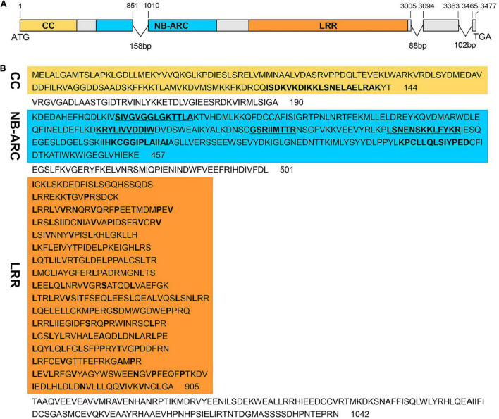
FIGURE 2.
Structure of RppM and its predicted protein domain and motifs. (A) Gene structure of RppM. The boxes represent exons, and lines denote introns. ATG and TGA are the translation start and stop codons, respectively. Yellow, blue, and orange regions represent the coiled-coil (CC) coding region, nucleotide-binding site (NBS), and the leucine-rich repeat (LRR) domain, respectively. (B) The predicted domain and motifs of RppM. The CC motif is indicated in bold font in the yellow region. The NBS domain is shown in blue, and the six conserved motifs (P-loop, Kinase2, RNBS-B, RNBS-C, GLPL, and MHDV) of the NBS are indicated in bold font. The C-terminal LRR domain, shown in orange, contains 17 LRR motifs.

