FIGURE 3.
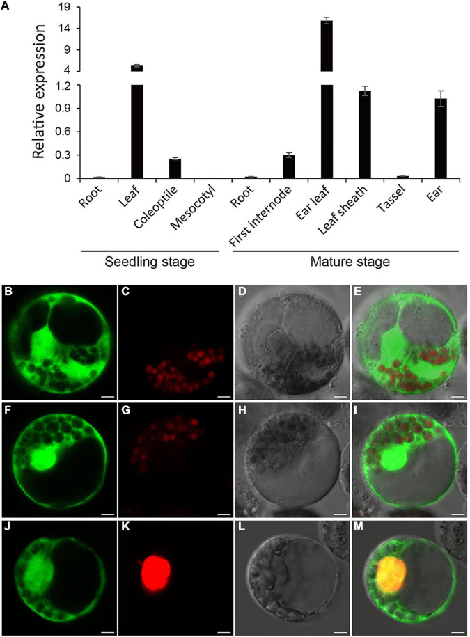
Expression pattern and of Subcellular localization of RppM. (A) Expression analysis of RppM in Jing2416K roots, leaves, coleoptiles, and mesocotyls at the seedling stage and roots, first internodes, leaf sheaths, tassels, and ears at the mature stage was performed by quantitative real-time PCR. Data are means ± SD of three biological replicates. (B–E) The free GFP. (F–M) Subcellular localization of RppM protein in maize protoplasts. (F,J) RppM-GFP fusion protein. (C,G) Chloroplast autofluorenscence. (K) The nucleus marker D53-mCherry. (D,H,L) Bright field image. (E) Merged image of (B,C). (I) Merged image of (F,G). (M) Merged image of (F,G). Scale bar: 5 μm.

