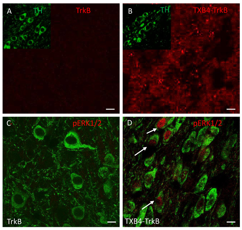
Figure 4.
Localization of human IgG, TH and pERK1/2 in the SNc of animals treated with TrkB agonist antibody or TXB4-TrkB. TrkB agonist antibody or TXB4-TrkB were administered SC to mice (10 mg/kg) and brains harvested after 18 h and stained (see methods for details). Within the SNc human IgG (red) was not detected after TrkB agonist antibody administration (A) but diffuse and punctate staining was seen following TXB4-TrkB administration (B). The inserts in both show dopaminergic neurons within the same field revealed by co-staining for TH. The same regions as in A and B were also co-stained for TH and pERK1/2. pERK1/2 was not detected at high magnification (63×) following TrkB agonist antibody administration (C) but was readily detected in some cell nuclei (as indicated by arrows) following TXB4-TrkB administration (D). Scale bars = 10 µm.

