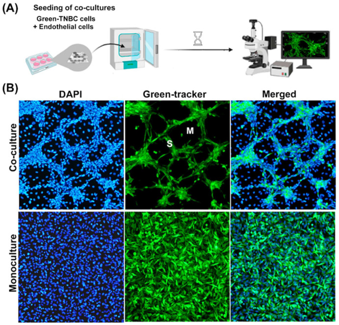
Figure 1.
Characterization of the co-culture model of TNBC cells with endothelial cells. (A) Illustration of the vasculogenic mimicry process in vitro. TNBC cells stained with a green tracker were seeded with endothelial cells (1:1) and allowed to interact for 2 days. The formation of cord-like networks was evaluated with epifluorescence microscopy. (B) MBCDF-T cells stained with a green cell fluorescent tracker were incubated in the presence (upper panels, co-cultures) or the absence (lower panels, monocultures) of unstained endothelial cells (EA.hy926). Afterwards, cells were fixed with ethanol. A mounting medium containing DAPI was used to label cell nuclei in blue (left panels). TNBC cells- vasculogenic mimicry was identified only in co-cultures by the formation of meshes (M) and segments (S) stained in green (upper panels). Merged pictures are shown in right panels. Magnification is 10×.

