Figure 2.
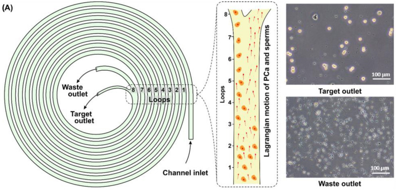
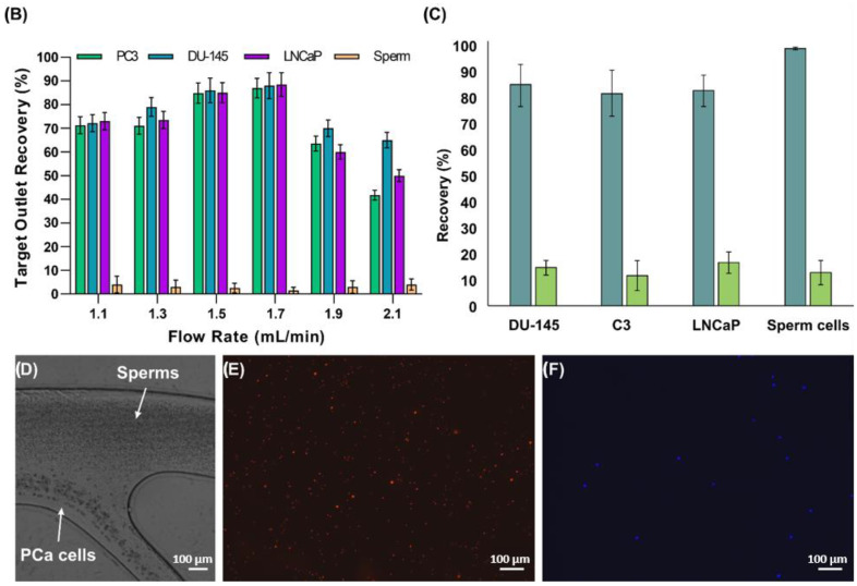
(A) Schematic representation of the spiral microfluidic channel for isolating CTCs from the SF. First, a preliminary prepared SF sample from a PCa patient is provided into the microchannel through the inlet under a continuous fluid flow at 1.7 mL/min. As a result, the SF sample is divided into target fraction containing most of the tumor cells and waste fraction containing most of the sperm cells. (B) Recovery rates of spiked PC3, DU-145, LNCaP cells, and sperm cells at different flow rates. (C) Recovery rates of spiked PCa tumor cells from three different cell lines (DU-145, PC3, and LNCaP) and sperm cells, after first and second runs of the device. The first run of the device implied the initial processing of the SF sample through the microfluidic chip. Thus, after the first run, it was possible to isolate more than 82% on average of the tumor cells, while more than 99% of the sperm cells could have been removed. The second run of the device implied processing of the target fraction. The processing of the target fraction resulted in a loss of the tumor cells and removal of the sperm cells in comparable percentages, at an average of 15% from the total number of tumor and sperm cells presenting in the target fraction. (D) High-speed camera image of CTC isolation from SF at the bifurcation point of the microfluidic channel. (E,F) Fluorescent images of target and waste fractions, respectively, showing a high concentration of DU-145 cells (blue dots) in the target fraction, and primarily sperm cells (red dots) in the waste fraction.


