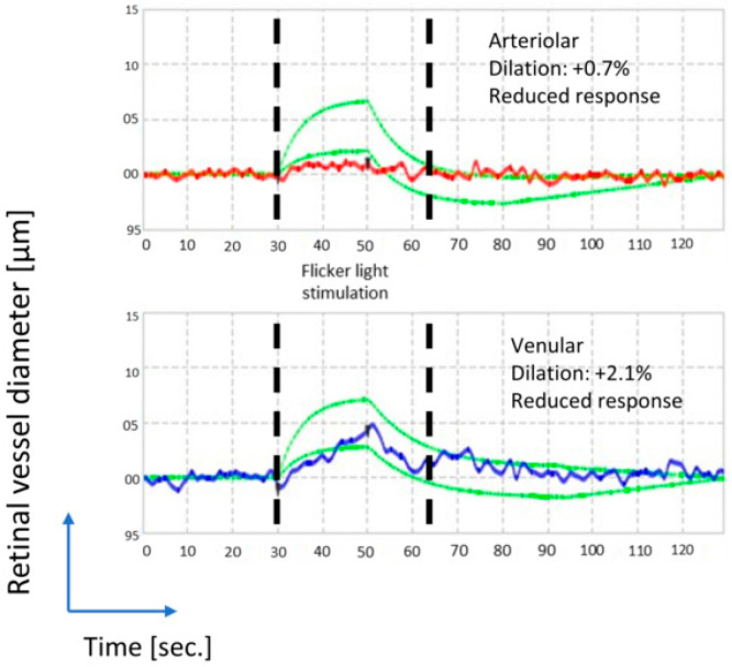
Figure 2.
Represents an example of vessel responsiveness measurements of a glaucoma patient to FL, using a retinal vessel analyser (RVA). On the y-axis the diameter (µm) of the selected vessel is plotted, and on the x-axis, the measurement during one period of flicker stimulation (sec). Retinal vessel responses to FL in the preselected vessel parts in contraction and in dilatation (%) were evaluated. Labelled in red (on the top) is the arterial and in blue (on the bottom), the venular diameter variation. The dashed green lines label the normal range. Both the AFR and the VFR are attenuated in glaucoma eyes, as exemplified here.

