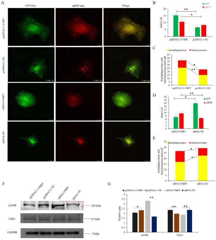
Figure 10.
miR-124a regulates myoblast autophagy via the transient receptor potential of the VMP1/ULK1 pathway: (A) An adenovirus harboring tandem fluorescent mRFP-GFP-LC3 was used to evaluate the extent of autophagic flux after overexpression or inhibition of miR-124a. (B,D) Mean numbers of GFP and mRFP puncta per cell; three cells were randomly selected from each field to be counted. (C,E) Mean numbers of autophagosomes and autolysosomes per cell. Autophagosomes have green and red puncta, while in the merged images the puncta appear yellow. Autolysosomes have red puncta only. (F,G) Protein levels of ULK1/mTOR were detected after overexpression or inhibition of miR-124a. GAPDH was used as an internal standard. Replicates = 3. Data are presented as the mean ± SE; * p < 0.05 and ** p < 0.01.

