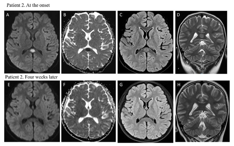
Figure 2.
MRI performed at the onset for patient 2 showed focal high signal intensity of splenium of the corpus callosum in diffusion-weighted images (DWI) (A) and low signal in ADC map (B) in the same lesion. Axial FLAIR (C) and coronal T2 w (D) images confirmed the presence of a hyperintense splenial lesion. MRI performed one month later revealed complete resolution of the lesion and normal signal intensity of splenium of corpus callosum (E–H).

