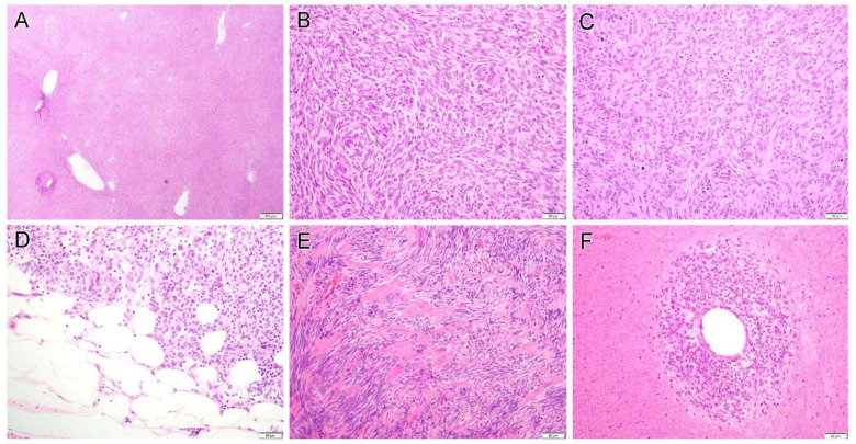
Figure 3.
Gastrointestinal stromal tumor displays a variety of growth pattern including hemangiopericytoma-like pattern (A, case 9), storiform (B, case 9) and haphazard (C, case 10) tumor growth, and fat involvement (D, case 13) can be seen in some areas. Some tumors contained hyalinized collagens (E, case 11). In some cases, viable cells are present only around the blood vessel and ghost-like outlines of necrotic atypical tumor cells can still be discerned in the surrounding tissue (F, case 16). Original magnification: (A), ×20; (B–F), ×200.

