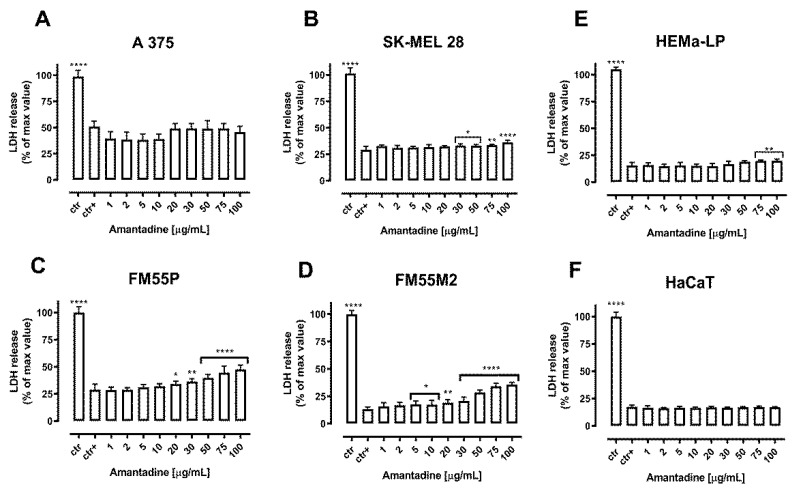
Figure 3.
Cytotoxicity of amantadine to malignant melanoma cells (A375 (A), SK-MEL28 (B), FM55P (C), FM55M2 (D)), melanocytes (HEMa-LP (E)) and keratinocyte (HaCaT (F)) measured by LDH assay. Results are presented as the percentage in LDH release to the medium by treated cells versus cells grown in control medium (ctr+) and cells treated with Lysis buffer (ctr). Data are presented as mean ± S.E.M. * p < 0.05, ** p < 0.01, **** p < 0.0001.

